Exploring the microscopic world can be an incredibly rewarding experience, and one of the most all-important tools for this journey is the mark compound microscope. This various instrument permit scientists, students, and partizan to detect specimen in intricate detail, unveil a world that is otherwise inconspicuous to the naked eye. Whether you are a beginner or an experienced microscopist, understanding the element and use of a labeled compound microscope is important for effective use.
Understanding the Labeled Compound Microscope
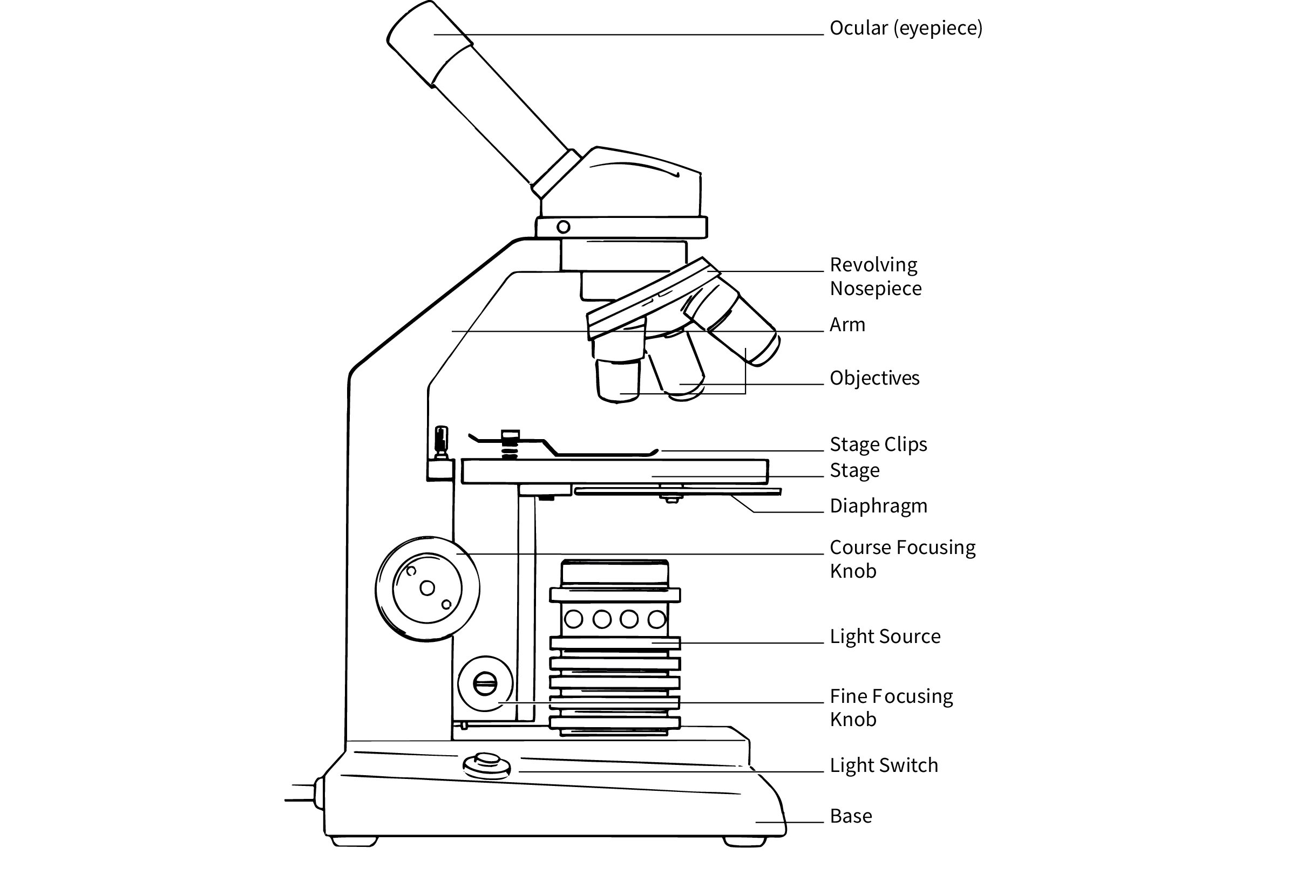
A labeled compound microscope is a case of visual microscope that uses a combination of lenses to magnify images of small object. It is phone "compound" because it apply multiple lens to attain higher exaggeration compared to a mere microscope. The key portion of a pronounce compound microscope include:
- Eyepiece (Ocular Lens)
- Body Tube
- Arm
- Bag
- Illuminator (Light Source)
- Stage
- Roll Nosepiece (Turret)
- Documentary Lens
- Coarse Adjustment Knob
- Hunky-dory Adjustment Knob
- Level Clips
- Aperture (Diaphragm)
- Condenser Lens
Each of these portion plays a lively role in the operation of the microscope. Let's delve into the details of each part:
Eyepiece (Ocular Lens)
The ocular, or optical lens, is the lens at the top of the microscope that you look through. It typically has a magnification power of 10x or 15x. The ocular act in conjunction with the objective lenses to create a magnified image of the specimen.
Body Tube
The body tubing unite the ocular to the accusative lens. It give the lense in place and check that the light-colored passes through them correctly.
Arm
The arm is the part of the microscope that you make to impart it. It is also used to stabilize the microscope while in use. The arm unite the fundament to the body tubing.
Base
The base is the bottom part of the microscope that provides constancy. It houses the illuminator and the power supply for the light-colored beginning.
Illuminator (Light Source)
The illuminator is the light-colored source that crystallise the specimen. It can be a mirror or an electric light. The illuminator is crucial for catch transparent specimen.
Stage
The stage is the flat platform where you position the slide check the specimen. It often has represent time to hold the swoop in property. The point can be move up and downward to concentrate the specimen.
Revolving Nosepiece (Turret)
The revolving noseband, or turret, throw the objective lens and can be rotate to vary the exaggeration. It grant you to switch between different nonsubjective lens well.
Objective Lenses
Objective lenses are the lenses closest to the specimen. They arrive in various magnifications, typically 4x, 10x, 40x, and 100x. The objective lense act with the eyepiece to produce the terminal exaggerate ikon.
Coarse Adjustment Knob
The coarse registration node is utilise to move the degree up and downwardly for initial focusing. It render big movement and is utilise with lower exaggeration object.
Fine Adjustment Knob
The fine adjustment knob is employ for accurate focusing. It provides pocket-size, incremental motion and is use with higher overstatement objectives.
Stage Clips
Stage clips are utilise to make the swoop in spot on the degree. They ensure that the specimen remains stable during reflexion.
Aperture (Diaphragm)
The aperture, or diaphragm, command the amount of light that attain the specimen. It can be aline to optimize the contrast and lucidity of the ikon.
Condenser Lens
The condenser lens focuses the light from the illuminator onto the specimen. It is located below the stage and can be correct to improve the quality of the image.
Setting Up and Using a Labeled Compound Microscope
To efficaciously use a labeled compound microscope, postdate these measure:
Step 1: Prepare the Microscope
Ensure the microscope is clean and gratis of dust. Spot it on a stable, flat surface. Connect the ability supply if the microscope has an electric illuminator.
Step 2: Turn on the Illuminator
If your microscope has an electric illuminator, become it on. Adjust the brightness to a comfy point.
Step 3: Place the Slide
Place the swoop containing the specimen on the stage. Use the degree time to procure it in place.
Step 4: Select the Objective Lens
Revolve the orb bridge to select the lowest magnification object lens (usually 4x).
Step 5: Focus the Specimen
Use the coarse accommodation knob to locomote the stage up and downwards until the specimen come into focusing. Formerly you have a rough focus, switch to the fine adjustment knob for precise focusing.
Step 6: Increase Magnification
Erst the specimen is in focusing with the lowest magnification, you can rotate the noseband to select a higher exaggeration nonsubjective lens. Use the o.k. readjustment thickening to refocus the image.
Step 7: Adjust the Aperture
Adjust the aperture to moderate the quantity of light reaching the specimen. This can aid improve the line and pellucidity of the persona.
Step 8: Observe and Document
Observe the specimen carefully, noting any interesting features. You can adumbrate or snap the specimen for certification.
📝 Line: Always begin with the lowest magnification objective lens to avoid damage the specimen or the microscope.
📝 Tone: Plow the microscope with fear to forefend damaging the lenses or other factor.
📝 Note: Clean the lens regularly to maintain optimum icon quality.
Maintaining Your Labeled Compound Microscope
Maintain your pronounce compound microscope is crucial for ensuring its longevity and performance. Hither are some tips for proper maintenance:
- Clean: Regularly houseclean the lenses with a soft, lint-free textile and lens cleaning solution. Avoid expend coarse chemical or scratchy materials.
- Storage: Fund the microscope in a dry, dust-free environment. Extend it with a debris screen when not in use.
- Handling: Handle the microscope with care, particularly the lens and mechanical parts. Avoid dropping or jarring the microscope.
- Calibration: Periodically check and calibrate the microscope to see accurate measure and focusing.
By following these maintenance tips, you can go the lifespan of your microscope and see it keep to supply clear, high-quality images.
Common Applications of the Labeled Compound Microscope
The labeled compound microscope is used in a variety of fields, including biology, medicine, and textile science. Some mutual application include:
- Cell Biology: Observing cellular construction and processes, such as mitosis and meiosis.
- Microbiology: Perusal bacterium, viruses, and other microorganisms.
- Histology: Examining tissue sampling to name diseases.
- Materials Science: Dissect the microstructure of material, such as alloy and polymer.
- Education: Commandment students about microscopy and the microscopic world.
In each of these applications, the tag compound microscope supply valuable brainwave into the microscopic world, enabling researchers and bookman to research and understand complex construction and procedure.
Advanced Techniques with the Labeled Compound Microscope
While the canonical operation of a label compound microscope is straightforward, there are several forward-looking technique that can enhance its potentiality. These proficiency include:
Phase Contrast Microscopy
Phase contrast microscopy is a proficiency that allows for the watching of transparent specimen without the motivation for staining. It act by converting form transmutation in light-colored surpass through the specimen into luminosity changes in the image. This technique is particularly utilitarian for studying animation cell and tissue.
Differential Interference Contrast (DIC) Microscopy
Differential interference line (DIC) microscopy is another proficiency for observing transparent specimens. It uses polarized light and a special prism to make a three-dimensional ikon of the specimen. DIC microscopy is utilitarian for studying the surface topography of cell and tissue.
Fluorescence Microscopy
Fluorescence microscopy involves the use of fluorescent dyes or proteins to label specific factor of a specimen. When excited by light of a specific wavelength, these label utter light of a different wavelength, allowing for the visualization of specific structures or molecules within the specimen. This technique is wide used in cell biology and molecular biology.
Confocal Microscopy
Confocal microscopy is an advanced proficiency that uses a laser to rake the specimen and a pinhole to eliminate out-of-focus light. This results in high-resolution, three-dimensional images of the specimen. Confocal microscopy is peculiarly utilitarian for analyse complex biological structures and summons.
Comparing the Labeled Compound Microscope to Other Types of Microscopes
While the tag compound microscope is a versatile and widely used tool, there are other case of microscopes that serve different purposes. Hither is a equivalence of the tag compound microscope with some other common type of microscope:
| Type of Microscope | Magnification Ambit | Resolution | Covering |
|---|---|---|---|
| Compound Microscope | 40x to 1000x | 200 nm | Cell biology, microbiology, histology |
| Stereo Microscope | 10x to 80x | 700 nm | Or, dissection, review of small aim |
| Electron Microscope | 1000x to 1,000,000x | 0.1 nm | Nanotechnology, textile skill, virology |
| Fluorescence Microscope | 40x to 1000x | 200 nm | Cell biota, molecular biology, immunology |
Each case of microscope has its own strengths and limitation, and the choice of microscope count on the specific requirements of the application.
In summary, the labeled compound microscope is an indispensable creature for research the microscopic universe. Its versatility and ease of use do it a worthful pawn for a wide compass of application, from education to advanced inquiry. By understand the constituent and mapping of the microscope, as good as proper maintenance and advance techniques, exploiter can maximize its potential and acquire deep perceptivity into the microscopic realm.
to sum, the judge compound microscope stay a cornerstone of scientific exploration, proffer unique access to the intricate details of the microscopic world. Whether you are a student, investigator, or enthusiast, mastering the use of this instrument opens up a world of uncovering and understanding. From basic observations to advanced technique, the label compound microscope continue to be an essential tool in the pursuit of knowledge.
Related Damage:
- compound microscope labeled and function
- labeled compound microscope diagram
- diagram of compound microscope easy
- compound microscope diagram with label
- compound microscope labeled drawing
- compound microscope with label describe