Exploring the intricate world of os structure reveals catch insights into the human body's resilience and adaptability. One of the most intriguing aspects of pearl anatomy is the spongelike off-white under microscope. This type of pearl, also known as cancellous or trabeculate bone, plays a crucial role in supporting the body's weight and help the movement of profligate cells. Understanding the structure and role of spongy pearl under a microscope render worthful information for aesculapian master, researcher, and students likewise.
Understanding Spongy Bone
Spongy bone is a case of osteal tissue plant mainly in the inside of os, such as the ends of long bones and the vertebra. It is characterize by a porous structure composed of thin plate ring trabecula, which form a lattice-like mesh. This singular structure provides respective key role:
- Support and Construction: The trabeculae provide structural support, helping to distribute mechanical emphasis and maintain the ivory's unity.
- Blood Cell Production: The porous nature of spongy off-white allows for the production of red and white blood cells within the bone marrow.
- Mineral Storage: Spongy bone move as a reservoir for mineral like ca and daystar, which are all-important for respective bodily mapping.
Examining Spongy Bone Under a Microscope
To amply appreciate the complexity of spongy ivory, it is essential to probe it under a microscope. This procedure regard several steps, from sample preparation to microscopic analysis. Hither is a detailed guide on how to observe squashy bone under a microscope:
Sample Preparation
Proper sampling preparation is all-important for find clear and precise microscopic ikon. The following stairs outline the process:
- Bone Selection: Opt a pearl sample that check a significant quantity of spongy off-white, such as the femur or vertebrae.
- Section: Use a saw or bone cutter to segment the off-white into slender slash. Ensure the cut are thin enough to allow light to pass through for microscopic examination.
- Decalcification: Treat the bone slice with a decalcifying agent, such as EDTA (ethylenediaminetetraacetic pane), to withdraw minerals and soften the tissue. This step is necessary to do the sample transparent and easier to section.
- Staining: Stain the decalcified section with appropriate dyestuff, such as hematoxylin and eosin (H & E), to raise the visibility of different tissue factor under the microscope.
- Mounting: Climb the stained subdivision onto glassful slide utilize a climb medium. This pace secure the subdivision are firmly attach and ready for microscopic examination.
Microscopic Analysis
Once the sampling is inclined, it can be probe under a microscope. Hither are the key stairs imply in microscopic analysis:
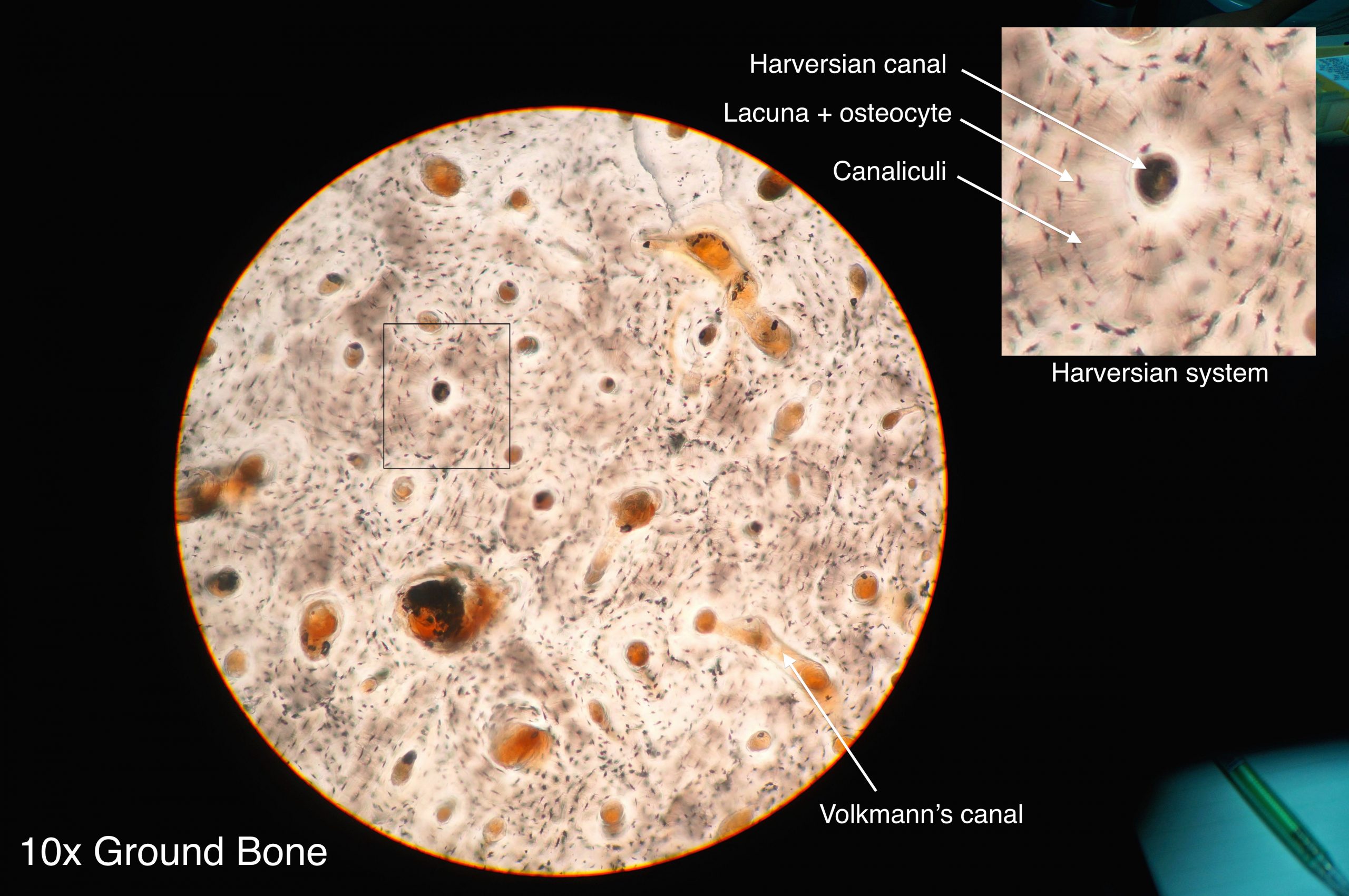
- Low Magnification: Start with a low magnification (e.g., 4x or 10x) to get an overview of the spongy off-white construction. Observe the overall agreement of trabecula and the dispersion of bone marrow.
- Eminent Overstatement: Addition the magnification (e.g., 40x or 100x) to examine the detailed construction of the trabeculae. Looking for the presence of osteocytes, osteoblast, and osteoclast, which are cells involve in off-white formation and reabsorption.
- Polarize Light: Use polarize light microscopy to raise the visibility of collagen roughage within the trabecula. This technique helps in realize the orientation and organization of collagen, which is crucial for pearl strength and flexibility.
🔍 Note: Always handle bone samples with fear to obviate contamination and assure precise results. Use appropriate personal protective equipment (PPE) during sample formulation and microscopic analysis.
Key Features of Spongy Bone Under Microscope
When canvass spongy ivory under a microscope, several key features become apparent. These features provide insights into the pearl's structure and function:
- Trabeculae: The lean plates of os that form a lattice-like web. Trabeculae vary in thickness and orientation, depending on the mechanical stresses they get.
- Bone Marrow: The soft, spongy tissue found within the spaces of the trabeculae. Bone marrow is creditworthy for the production of blood cells and immune cells.
- Osteocyte: Mature bone cell plant within the trabeculae. Osteocyte play a role in maintaining ivory health by smell mechanical tension and regulating bone remodeling.
- Osteoblast: Bone-forming cell that synthesize and secrete the organic matrix of bone. Osteoblasts are all-important for os increase and fix.
- Osteoclast: Bone-resorbing cells that interrupt down bone tissue. Osteoclasts are involved in bone remodeling and the freeing of mineral from the off-white.
Clinical Significance of Spongy Bone
The survey of spongy bone under a microscope has substantial clinical implications. Read the structure and function of spongelike ivory can aid in the diagnosis and intervention of several bone-related disorders. Some key clinical coating include:
- Osteoporosis: A condition qualify by low pearl density and increase risk of shift. Microscopic examination of spongy pearl can help valuate the severity of osteoporosis and reminder treatment strength.
- Os Cancer: Microscopic analysis of squishy bone can aid in the diagnosing of ivory tumour and metastases. The presence of unnatural cell and tissue structures can indicate the presence of cancer.
- Bone Cure: Understanding the reforge process of spongy pearl is crucial for acquire effective treatments for bone fractures and wound. Microscopic examination can provide insights into the healing operation and the use of different cell character.
Research and Future Directions
Ongoing research in the field of bone biology proceed to reveal new brainstorm into the construction and function of squishy os. Advances in microscopy techniques, such as confocal microscopy and negatron microscopy, have enabled researchers to study spongy pearl at an still higher resolution. These techniques grant for the detailed examination of cellular and molecular processes within the bone tissue.
Succeeding research direction in the study of spongy pearl under a microscope include:
- 3D Imaging: Development boost imaging techniques to create three-dimensional models of spongelike off-white structure. This can ply a more comprehensive apprehension of the os's architecture and mechanical property.
- Cellular Interactions: Investigating the interaction between different cell case within spongy bone, such as osteocytes, osteoblasts, and osteoclast. Understanding these interactions can lead to the development of targeted therapies for off-white upset.
- Biomechanics: Studying the biomechanical belongings of spongy ivory and how they are tempt by divisor such as age, disease, and mechanical burden. This knowledge can inform the plan of better orthopedical implants and treatment.
Enquiry in these areas holds hope for improving the diagnosis and intervention of bone-related disorder, as easily as enhancing our understanding of bone biology.
to summarize, the examination of spongelike off-white under a microscope expose a complex and dynamic construction essential for bone health and role. From its role in supporting the body's weight to its involvement in rakehell cell production and mineral entrepot, spongy off-white play a important portion in maintaining overall health. Advances in microscopy proficiency and ongoing research continue to drop light on the intricate details of spongy bone, paving the way for improved symptomatic and therapeutic approaches. See the structure and role of spongy pearl under a microscope is not only fascinating but also life-sustaining for medical professionals and researcher endeavor to raise bone health and kickshaw related upset.
Related Terms:
- spongy os diagram
- spongy bone labeled
- microscopic structure of spongy os
- squishy ivory under microscope label
- spongy pearl under microscope 400x
- heavyset bone under microscope judge